Indiana Microscopy Society
An Affiliate of the Microscopy
Society of America

|

|

|

|
 |

|
 |

|

|
|
|
Indiana Microscopy SocietyAn Affiliate of the Microscopy
|
|
Dear Members, Corporate Sponsors and Students,
The INMS Spring Meeting will be held Friday, May 11, 2012, at Eli Lilly Corporate Center. Our host at Lilly is Jeff Hanson. Thanks to Jeff for clearing this with his management and making arrangements for lunch, meeting space and parking. Lunch is free for all paid members.
Important note: For entry into Lilly buildings, you must have a picture ID – a driver’s license is acceptable. If you are planning to attend please, email Mike Esterman that you are coming and the number and names of the persons in your group if you are responding for more than yourself.
If you are not sure if your dues are paid, I will check and send you an email confirming your reservation and your dues status. If you haven’t paid for 2012, please remit $10 to INMS and send to Mike Esterman, Secretary, 7263 North Baltimore Rd, Monrovia, IN 46157.
Registration will begin at 9 am, and presentations will run from 10 am until 4pm with a lunch from noon until 1:30 pm. We extended the lunch time to give you an opportunity to eat, visit with other members and visit the Corporate Sponsors Tables.
Directions to the Lilly Corporate Center , parking, speakers, titles, abstracts and the schedule will be sent in a separate email notice in about 10 days.
The speakers will provide a diverse and informative look at non-traditional imaging methods:
John Sullivan, Lilly Research Labs, Welcome
Bogdan Dragnea, Department of Chemistry, Indiana University, "Photothermal imaging of virus like particles: a possible alternative to single particle tracking by fluorescence microscopy"
Steven L. Tait, Department of Chemistry, Indiana University, "Scanning Probe Microscopy: Precision Imaging of Atomic and Molecular Ordering at Surfaces"
Brandon Hirsch, Department of Chemistry, Indiana University, "Self-Assembly of Aryl-Triazole Oligomers at the Liquid/HOPG Interface imaged by Scanning Tunneling Microscopy"
Steven L. Goodman, Microscopy Innovations, LLC, "A Better Way to Prepare and Handle Microscopy Specimens and TEM Grids"
Donald A. Chernoff, Advanced Surface Microscopy Inc., “Atomic Force Microscopy - from Nanomedicine to Consumer Products”
Check out the newest addition to the INMS website, our gallery of member images. The gallery now has its first set of images. If you have images to contribute to the gallery page, you may send them to Mike Esterman using either e-mail (if not too many or too large), a CD or DVD or we can set up a place to deposit them. E-mail Mike Esterman about contributing images to the gallery.
The purpose of these member profiles is to help the INMS membership learn the scientific and personal interests of various members to foster networking within our society.
Corporate Sponsor Member Bill Chauvin grew up in Madison, WI. Bill’s interest to pursue microscopy science grew from initial interest in pre-medicine which turned into a pursuit of cell biology/ultrastructure. He obtained his education in General Science from University of Western Ontario were he also earned a MSc in Zoology-Cell Biology. Bill is currently employed by Corporate Sponsor, Oxford Instruments. He has extensive experience with many models of SEM and TEM originally for cell transport mechanisms and Pathology, and eventually for semiconductor and materials research. Bill has employed Cameca IMS 3F SIMS for semiconductor research and geological research, XPS for semiconductors and catalysis and PHI Auger for semiconductors and corrosion. His hobbies include golf and duplicate bridge.
Bill’s advice to fellow microscopists is “pursue microscopy like astronomy. Most of the infinitely small universe is as unexplored as the infinitely vast.”
Corporate Sponsor Member Jerry Jasso grew up in Grand Rapids, MI. Jerry was led to pursue science as a career by his life sciences professor who provided great encouragement to pursue his career goals in Microbiology. Jerry earned a BSc in Microbiology and an MBA. He is currently employed by Corporate Sponsor, Ted Pella, Inc. Jerry has enjoyed a career that has embraced several modes of imaging and microscopy: his career profile includes many years of EM in both Research and Clinical applications and also, IHC/Molecular Diagnostics, Histology, Flow Cytometry, and Confocal Microscopy.
Jerry says, “Ask me about the 'Smerf Hog'” So the next time you need to confer with Jerry, find out what that means! Jerry’s advice to fellow INMS members is to be aware that as life and material sciences meld through nanotechnology, the field of microscopy has become increasingly important as a research tool, thus investigators must fully understand this tool’s evolving capabilities to meet today’s research challenges.
Expand your knowledge of microscopy with MSA membership! Regular membership is $60 per year and Student membership is $20 per year. Members receive: subscription to Microscopy and Microanalysis and Microscopy Today, discounts on books, journals and other educational materials, MSA awards programs, speaker opportunities, and scholarships, and peer networking through the society's focused interest groups. The Microscopy and Microanalysis 2013 conference will be right here in Indianapolis.
Dr. Brad Amos, MRC Laboratory of Molecular Biology, Cambridge, UK, is developing a giant optical lens to provide high resolution optical sectioning over a large specimen area (6 mm diameter). Read about this development or listen to Brad talk about it (when the new page comes up, scroll down just a little and look on the left hand side list of lectures for "How new science is transforming the optical microscope, Dr Brad Amos FRS"; when last checked, it was the 3rd lecture listed from the top).
There is also an interesting web article from Zeiss on the effect of coverslip thickness on image quality.
The Generation 6 XFlash SDD detectors have active areas of 10, 30, 60 and 100 sq mm, are available for SEM and TEM, and support count rates of 1.5 million cps. For information contact Mark Kelsey.
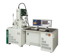
The new JEOL JSM-7100F FE SEM offers expanded performance to the budget-conscious lab, featuring 10x-1,000,000x magnification and analytical resolution at the sub 100 nm scale. When equipped with low vacuum (LV) mode (up to 300 Pa), all LV orifices can be retracted without breaking vacuum for unrestricted low magnification imaging and maximum beam current (200 nA) in high vacuum. For the ultimate versatility, the JSM-7100FTLV features through-the-lens electron detectors with energy filter. Check out the corporate website or or e-mail their sales force.
The TEAM™ Pegasus system combines world-class Energy Dispersive Spectroscopy (EDS) and Electron Backscatter Diffraction (EBSD) hardware with EDAX’s highly successful TEAM™ software platform to create the next generation in synergistic materials characterization.
TEAM™ Pegasus is the integration of EDAX’s TEAM™ EDS analysis system and its market-leading EBSD product line. The product offers both high-level analysis capabilities and an easy-to-use, intuitive interface to users of all levels. The TEAM™ platform introduces Smart Features to EBSD. Smart Camera and Smart Background guide the user in optimal data collection, and Smart Indexing and Smart Data Management guarantee accurate analysis and reporting. Look here for a complete description of TEAM™ Pegasus
The mPrep™ Capsule Grid Box offers secure storage of TEM grids within individually labeled capsules. Provided free-of-charge with purchase of mPrep/g capsules, the box can hold 32 grids in 16 capsules. Patented mPrep/g capsules provide an enclosed environment for grid-staining, which reduces specimen “touches” and reagent consumption. Capsule processing also enables barcode or alphanumeric labeling of valuable grids. Resin blocks prepared in soon-to-be-released mPrep/s capsules can be stored together in the same box with their corresponding grids, offering a convenient way to keep an entire project together.
For more information, visit the corporate website or call toll-free 888-302-3925.
| Applied Precision www.api.com |
Bruker www.bruker.at |
Carl Zeiss Microscopy www.zeiss.com/nts |
EDAX www.edax.com |
| Electron Microscopy Sciences www.emsdiasum.com |
FEI Company www.fei.com |
Gatan, Inc. www.gatan.com |
Hitachi HTA www.hitachi-hta.com |
| IXRF Systems, Inc www.ixrfsystems.com |
Mager Scientific, Inc www.magersci.com |
Microscopy Innovations www.microscopyinnovations.com |
Oxford Instruments www.oxford-instruments.com |
| Ted Pella, Inc www.tedpella.com |
Mike Esterman, Secretary
Indiana Microscopy Society
7263 North Baltimore Rd
Monrovia, IN 46157
esterman@ccrtc.com