We are just beginning to collect images from INMS members. Here are some images sent to us by our Secretary, Mike Esterman:
|
|
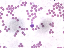
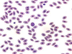
Blood cells. Human red blood cells (left panel) and avian red blood cells (right panel) prepared with Wright's stain. The avian red blood cells are nucleated and oval while the human red blood cells lack nuclei and appear circular. Both bright field images were recorded at 63x. |
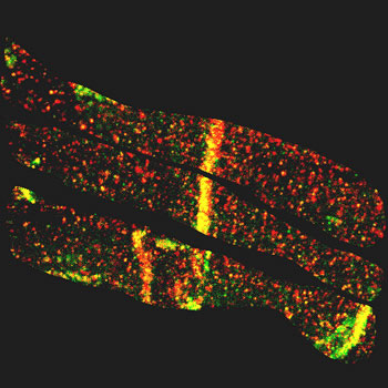 |
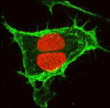
Mouse cardiac muscle. In this confocal image, connexin 43 is stained green and PKC-alpha is stained red. These two proteins co-localize strongly in the interculated disk (the communication junction between heart cells) as seen in the regions of this image that appear yellow. |
|
|
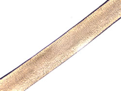
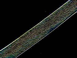
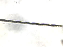
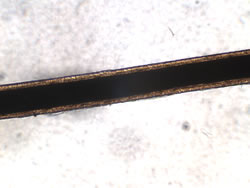
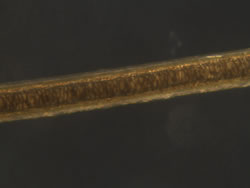
Hair. All these samples were embedded in glycerol which gives high contrast due to its refractive index. The top pair of images shows a bright field (left) and dark field (right) image of a human hair. The BF image was processed using both background subtraction and some contrast enhancement to reveal the scales (ridge-like texture). The DF image was treated with a horizontal edge filter to reveal the texture. The middle pair of images are bright field images of horse (left) and cat (right) hairs. This type of horse hair has a very narrow, dark core (compare to the BF image of the dog hair immediately below). The cat hair is much narrower than the other types of hair shown here and appears to be segmented along its length. The bottom pair of images are bright field (left) and dark field (right) images of a dog hair. The BF image shows the very large, dark core typical of non-human species. The DF image allows for more detail to be seen in the core region. |
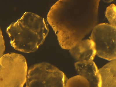 |
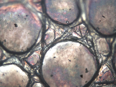
Sand. Dark field image (10x) of sand used for making concrete. Dark field imaging is a good choice when dealing with opaque samples such as this. |
|
|
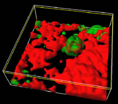
Confocal images. 20x image of blood vessels in a mouse solid tumor revealed by IV injection of a fluorescently labeled high molecular weight dextran (left) and a 40x image of HeLa cells labeled with propidium iodide (red staining in nuclei) and an Alexa 488 stained transporter protein in the plasma membranes (green staining). |
 |
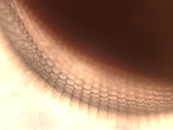
Lizard skin. Bright field image recorded at 10x of lizard skin. |
|
|
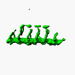
Three-dimensional (3d) models produced from confocal images. Top image pair: 3d models of blood vessels in a live zebra fish where the endothelial cells have been labeled with GFP (left) and a brain astrocyte assimulating A-beta protein (one of the proteins implicated in Alzheimer's Disease). Bottom image: 3d model of HeLa cell mitotic spindles showing how cells in mitosis stand up from the growing surface. An anti-cancer drug that inhibits mitosis was used to prevent the mitotic spindles from separating the chromosomes into daughter cells. |
|
|
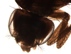
Fruit fly images. The left panel shows an entire fly head including the faceted eyes and antenna along with the front legs and part of the fly body (bright field image at 10x). The right panel is a close-up of the eye and shows hair-like structures between the eye facets (bright field image at 40x). |
|
|
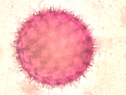
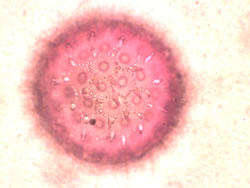
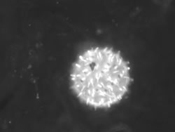
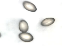
Pollen. The top panel shows two bright field images of the same morning glory pollen grain recorded at 63x. The images show different focal planes. The spikes seen clearly in the image on the left are sticky and facilitate pollen sticking to insects which helps in pollenation. Pore-like structures are clearly seen in the second image (right). The bottom panel shows an okra pollen grain (left) and several camelia pollen grains (right). The okra pollen fluorescence image is the only image in this set recorded at 20x and shows how much larger (>200 microns) these pollen grains are relative to the others. This particular pollen grain is auto-fluorescent. The bright field image of camelia pollen (recorded at 63x) reveals that these pollen grains are small, smooth and oval. Camelias are self-pollenating flowers and the smooth structure of their pollen prevents it from sticking to insect legs. |